Hypoxemic failure (type 1) shows PaO2 below 60; hypercapnic failure (type 2) shows PaCO2 above 50 with pH below 7.35.
Before you read
Test yourself first: 5 questions
Question 1 of 5
During an acute asthma attack, which assessment finding would most concern the nurse and suggest worsening airflow obstruction?
Answer: B. A suddenly quiet or silent chest during a severe asthma attack indicates airflow is so limited that air movement is minimal, a sign of impending respiratory failure. Loud wheezing, by contrast, means air is still moving. This is an emergency requiring immediate intervention.
Question 2 of 5
A 5-year-old is having an acute asthma exacerbation. Which assessment finding indicates the exacerbation is worsening and warrants immediate intervention?
Answer: B. A silent or markedly diminished chest with absent wheezing during an asthma attack signals severely reduced air movement and impending respiratory failure. It is more ominous than loud wheezing, which at least indicates air is moving.
Question 3 of 5
A client with acute decompensated heart failure presents with dyspnea, frothy pink-tinged sputum, and crackles throughout both lung fields. Which position should the nurse place the client in FIRST?
Answer: B. High Fowler position (sitting upright, often with legs dependent) reduces venous return (preload) and maximizes lung expansion, easing the work of breathing in acute pulmonary edema. Supine, Trendelenburg, and left lateral positions increase venous return to an already overloaded heart and worsen respiratory distress.
Question 4 of 5
A 6-month-old infant is admitted with bronchiolitis caused by respiratory syncytial virus (RSV). Which assessment finding should the nurse report to the provider as the priority concern?
Answer: C. Nasal flaring and intercostal retractions indicate increased work of breathing and impending respiratory failure, the priority in RSV bronchiolitis. Clear nasal drainage, a low-grade fever, and mild cough are expected, less acute findings.
Question 5 of 5
A client with myasthenia gravis is at risk for a myasthenic crisis, which causes:
Answer: B. Myasthenic crisis causes severe muscle weakness including the respiratory muscles → respiratory failure. Monitor respiratory status closely; it can be life-threatening.
Keep the streak going with more questions like these, each with a full rationale.
What is acute respiratory failure?
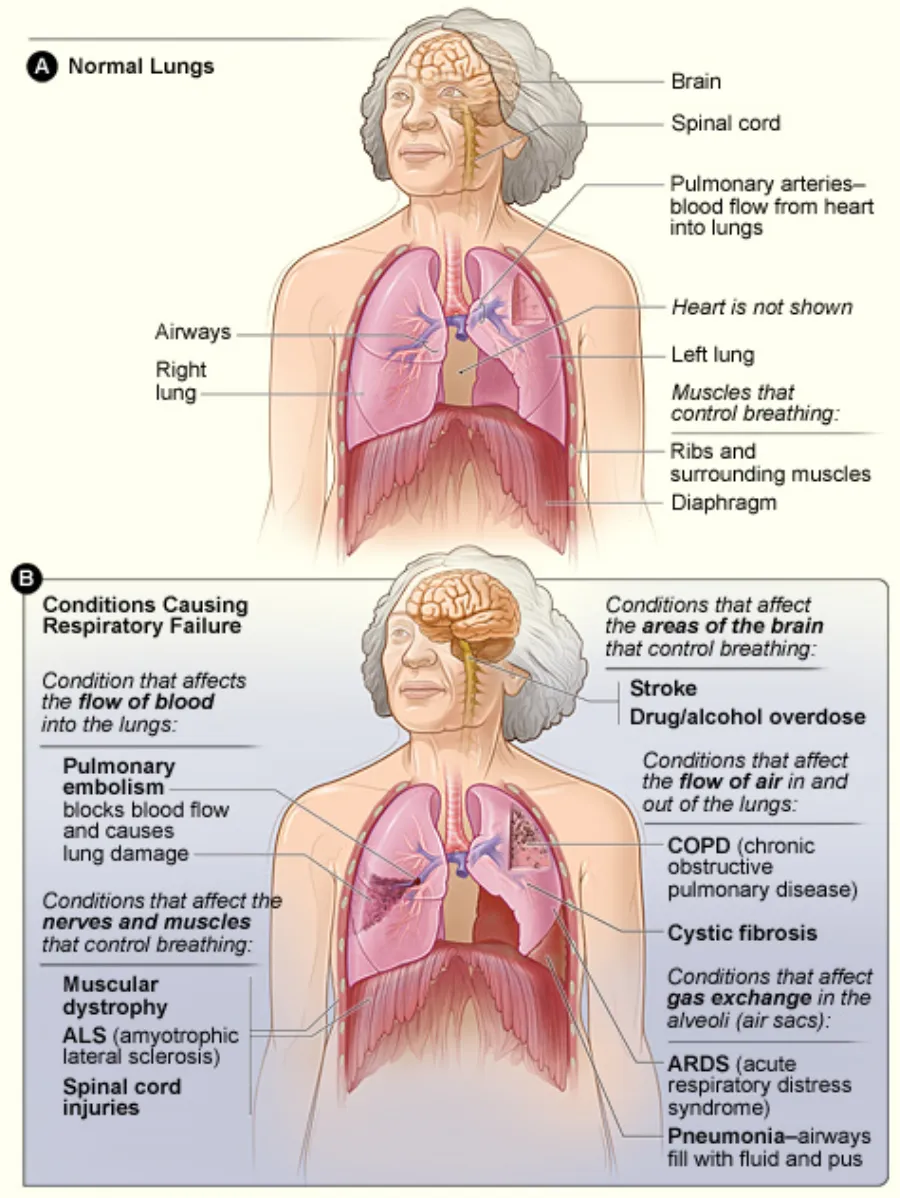
It is a medical emergency that can result from pneumonia, COPD exacerbation, PE, ARDS, or neuromuscular weakness. Early recognition of restlessness and the work of breathing is critical to prevent arrest.
Acute Respiratory Failure illustrated
Key points to remember
- Hypoxemic failure (type 1) shows PaO2 below 60; hypercapnic failure (type 2) shows PaCO2 above 50 with pH below 7.35.
- Early signs are restlessness, anxiety, confusion, and tachycardia from hypoxia.
- Dyspnea, tachypnea, accessory muscle use, and nasal flaring reflect increased work of breathing.
- Late signs include cyanosis, decreased level of consciousness, bradycardia, and a falling respiratory rate.
- Headache, drowsiness, and flushed skin suggest carbon dioxide retention.
- A decreasing respiratory rate with worsening hypoxia signals fatigue and impending arrest, not improvement.
Signs & symptoms
- Restless, confusion = early hypoxia
- Accessory muscles, retractions
- Cyanosis, ↓ LOC = late/severe
Nursing priorities
- Apply oxygen and position the patient upright to maximize ventilation.
- Maintain a patent airway and prepare for noninvasive ventilation or intubation if needed.
- Call the rapid response team and obtain ABGs to guide management.
- Treat the underlying cause, such as a bronchodilator for bronchospasm or antibiotics for infection.
- Continuously monitor oxygenation, ventilation, level of consciousness, and cardiac rhythm.
Labs & values
Red flags: report now
- A falling respiratory rate, rising CO2, and decreasing level of consciousness indicate impending respiratory arrest.
- Cyanosis, bradycardia, and unresponsiveness require immediate airway intervention.
- In a COPD patient, high-flow oxygen can suppress the hypoxic drive, so titrate to the lowest effective level and watch for rising CO2.
References
Clinical references for this topic