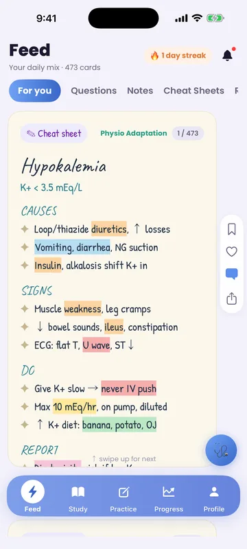
Earliest signs are muscle twitching/cramps progressing to weakness and flaccid paralysis
Before you read
Test yourself first: 3 questions
Question 1 of 3
A client has a serum potassium level of 6.8 mEq/L. Which ECG change should the nurse anticipate?
Answer: B. Hyperkalemia classically produces tall, peaked T waves. As levels rise further, the QRS widens and the P wave flattens. Prolonged QT and U waves are features of hypokalemia, not hyperkalemia.
Question 2 of 3
A client has a serum potassium of 6.8 mEq/L. Which finding on the cardiac monitor should the nurse expect?
Answer: A. Hyperkalemia classically produces tall, peaked T waves as one of the earliest ECG changes; widening QRS and bradydysrhythmias follow as it worsens. A shortened QT and prominent U waves are seen in other imbalances (hypercalcemia, hypokalemia), not hyperkalemia.
Question 3 of 3
A client reports muscle weakness and palpitations. The ECG shows a flattened T wave and a U wave, and labs are pending. Which electrolyte imbalance does the nurse suspect?
Answer: A. Flattened T waves, prominent U waves, ST depression, muscle weakness, and palpitations are classic for hypokalemia. Hyperkalemia shows peaked T waves; calcium and magnesium disturbances produce different ECG and neuromuscular patterns.
Keep the streak going with more questions like these, each with a full rationale.
What is hyperkalemia?
Because potassium controls cardiac membrane excitability, a high level is dangerous mainly because of life-threatening dysrhythmias. Common causes are renal failure, potassium-sparing diuretics, ACE inhibitors, Addison's disease, tissue breakdown (crush injury, burns), and acidosis shifting K⁺ out of cells.
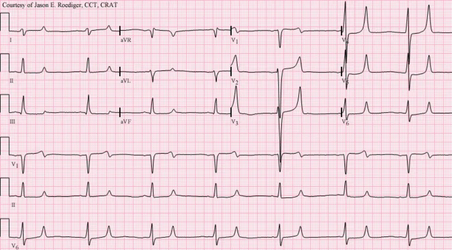
Hyperkalemia illustrated
Key points to remember
- Earliest signs are muscle twitching/cramps progressing to weakness and flaccid paralysis
- ECG changes: tall, peaked T waves, widened QRS, and prolonged PR — can progress to asystole
- GI: hyperactive bowel sounds, diarrhea, abdominal cramping
- Renal failure is the most common cause
Common causes
- Renal failure = #1 cause
- K-sparing diuretics, ACE, ARBs
- Acidosis, cell lysis, burns
Signs & symptoms
- Peaked T waves, wide QRS
- Muscle twitch → flaccid paralysis
- ↑ bowel sounds, diarrhea
Nursing priorities
- Cardiac monitor — peaked T waves signal emergent risk
- Calcium gluconate IV to stabilize the myocardium (does not lower K⁺)
- Shift K⁺ into cells: IV insulin with dextrose, and/or nebulized albuterol, sodium bicarb if acidotic
- Remove K⁺ from the body: sodium polystyrene sulfonate (Kayexalate) or dialysis
- Stop all potassium sources — IV fluids, supplements, K⁺-sparing diuretics
Labs & values
Patient teaching
- Renal clients: avoid salt substitutes (they are potassium chloride) and high-K⁺ foods
Red flags: report now
- K⁺ > 6.5 mEq/L or peaked T waves / widening QRS — treat as an emergency
- Hold ACE inhibitors and potassium-sparing diuretics (spironolactone)
References
Clinical references for this topic