Increasing ulcer size, foul drainage, fever, or surrounding redness indicates infection — report.
Before you read
Test yourself first: 4 questions
Question 1 of 4
A nurse assesses a client and identifies findings consistent with a venous stasis (venous insufficiency) leg ulcer. Which set of findings is most characteristic of a venous ulcer?
Answer: B. Venous insufficiency ulcers typically occur over the medial malleolus (inner ankle), have irregular borders, are shallow with moderate to heavy drainage, and are accompanied by edema, brownish (hemosiderin) skin discoloration, and warm skin with present pulses. Deep, pale, painful ulcers on the toes/lateral foot with absent pulses suggest arterial disease.
Question 2 of 4
A nurse is positioning a client with increased ICP. Which position best promotes venous drainage from the brain and helps lower ICP?
Answer: A. Head of bed elevated about 30 degrees with the head and neck in neutral, midline alignment promotes jugular venous outflow and lowers ICP. Flexing or rotating the neck, hip flexion, and the flat or Trendelenburg position impede venous drainage and raise ICP.
Question 3 of 4
A client with chronic pancreatitis is being evaluated for malabsorption. Which stool finding does the nurse expect?
Answer: B. Chronic pancreatitis reduces secretion of lipase and other digestive enzymes, leading to fat malabsorption and steatorrhea: bulky, greasy, foul-smelling, pale stools that float. Clay-colored stools suggest biliary obstruction, melena reflects upper GI bleeding, and bright red blood per rectum suggests a lower GI source. Greasy, foul-smelling fatty stools are the classic finding of pancreatic exocrine insufficiency.
Question 4 of 4
A nurse is assessing a client with chronic bronchitis, a type of COPD. Which finding is characteristic of this condition?
Answer: A. Chronic bronchitis is defined by a chronic productive cough with excessive mucus production for at least 3 months in 2 consecutive years; clients are often described as having cyanosis and edema. A thin body habitus with pursed-lip breathing and minimal sputum more typically describes emphysema. The other findings are not characteristic.
Keep the streak going with more questions like these, each with a full rationale.
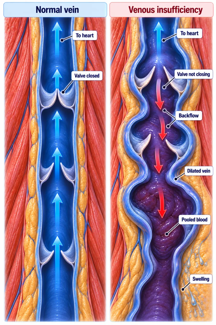
What is chronic venous insufficiency?
Blood pools in the lower legs, causing increased pressure and tissue damage. Venous (stasis) ulcers typically appear over the medial ankle.
Chronic Venous Insufficiency illustrated
Key points to remember
- Caused by chronic venous valve incompetence or prior DVT, leading to venous hypertension.
- Hallmarks: lower-leg edema, brownish hyperpigmentation, and stasis dermatitis.
- Venous stasis ulcers occur over the medial malleolus (inner ankle), are shallow with irregular edges and drainage.
- Legs are usually warm with present pulses and edema — unlike cool, pulseless arterial disease.
- Elevation and compression therapy are mainstays of management.
- Distinguish from arterial ulcers, which are deep, painful, punched-out, and on the toes/lateral foot.
Signs & symptoms
- Brown hemosiderin staining
- Edema, warm, +pulses
- Ulcers at medial malleolus
Nursing priorities
- Apply compression therapy (stockings or wraps) as prescribed to promote venous return.
- Elevate the legs above heart level frequently to reduce edema.
- Provide wound care for venous ulcers and monitor for infection.
- Inspect skin and protect it from breakdown and trauma.
- Confirm adequate arterial flow (ankle-brachial index) before applying compression.
Labs & values
Patient teaching
- Wear compression stockings daily and elevate the legs whenever resting.
- Inspect skin daily, keep it clean and moisturized, and avoid injury.
- Avoid prolonged standing or sitting and do not cross the legs.
- Report any new sores, increasing swelling, or signs of infection.
Red flags: report now
- Increasing ulcer size, foul drainage, fever, or surrounding redness indicates infection — report.
- Compression applied without verifying arterial flow can worsen arterial insufficiency.
- Sudden calf pain and swelling may signal a new DVT.
References
Clinical references for this topic