Central vision is lost or distorted while peripheral vision remains intact (opposite pattern to glaucoma).
Before you read
Test yourself first: 2 questions
Question 1 of 2
A nurse reviews the clinical picture of a client with amyotrophic lateral sclerosis (ALS). Which combination of findings is consistent with this disorder?
Answer: A. ALS is a progressive degeneration of both upper and lower motor neurons, producing muscle weakness, atrophy, fasciculations, and spasticity while sensation and cognition usually remain intact. Loss of sensation, relapsing-remitting vision changes, and a resting tremor are not characteristic of ALS.
Question 2 of 2
A nurse cares for a client with myasthenia gravis. Which statement best describes the underlying pathophysiology the nurse should understand?
Answer: B. Myasthenia gravis is an autoimmune disorder in which antibodies attack acetylcholine receptors at the neuromuscular junction, impairing nerve-to-muscle transmission and causing fluctuating, fatigable muscle weakness. It is not caused by dopamine deficiency, demyelination of central neurons, or motor neuron degeneration.
Keep the streak going with more questions like these, each with a full rationale.
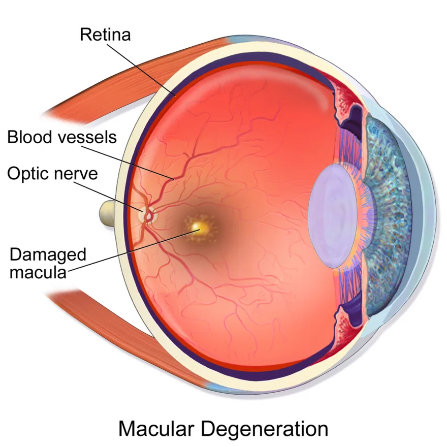
What is macular degeneration?
The dry (atrophic) form is most common and slowly progressive; the wet (exudative) form involves abnormal leaking blood vessels and causes more rapid, severe central vision loss. It is a leading cause of blindness in older adults. Patients lose the ability to read, recognize faces, and do fine detail work.
Macular Degeneration illustrated
Key points to remember
- Central vision is lost or distorted while peripheral vision remains intact (opposite pattern to glaucoma).
- An early sign is metamorphopsia: straight lines appear wavy or distorted, detected with an Amsler grid.
- A central blind spot (scotoma) develops, making reading and recognizing faces difficult.
- Dry AMD progresses slowly with drusen deposits; wet AMD causes sudden, rapid central vision loss from leaking vessels.
- Major risk factors are advancing age, smoking, family history, and Caucasian race.
Signs & symptoms
- Central vision loss, blurry
- Straight lines look wavy
- Amsler grid for monitoring
Nursing priorities
- Teach the patient to use an Amsler grid at home daily and report new distortion or worsening central vision.
- For wet AMD, prepare for and support intravitreal anti-VEGF injections that slow vessel growth.
- Promote low-vision aids (magnifiers, large-print, high-contrast lighting) and refer to vision rehabilitation.
- Encourage smoking cessation and a diet rich in leafy greens; reinforce AREDS antioxidant vitamins if prescribed.
- Modify the environment for safety and maximize use of remaining peripheral vision.
Patient teaching
- Check the Amsler grid one eye at a time daily and report any new wavy lines or blank spots.
- Stop smoking and eat dark leafy green vegetables; take prescribed eye vitamins.
- Use bright lighting, magnifiers, and large-print materials to make the most of remaining vision.
Red flags: report now
- Sudden worsening of central vision or new distortion may signal conversion to wet AMD; urgent ophthalmology referral.
- New flashes, floaters, or a curtain over vision suggest a different emergency (retinal detachment); escalate.
- Reinforce that vision loss is permanent so patients avoid unproven 'cures' and stay in treatment.
- Do not delay reporting changes; wet AMD treatment is time-sensitive.
References
Clinical references for this topic