Most common cause is rheumatic fever following untreated strep infection.
Before you read
Test yourself first: 3 questions
Question 1 of 3
A nurse auscultates a high-pitched, blowing systolic murmur heard best at the apex and radiating to the axilla in a client. This finding is MOST consistent with which valvular disorder?
Answer: B. Mitral regurgitation produces a holosystolic, blowing murmur heard best at the apex (fifth intercostal space, midclavicular line) that radiates to the left axilla. Aortic stenosis is a systolic murmur at the right second intercostal space radiating to the carotids; mitral stenosis and aortic regurgitation are diastolic murmurs.
Question 2 of 3
A client with carotid artery stenosis is scheduled for a carotid endarterectomy. Postoperatively, which assessment finding requires IMMEDIATE intervention?
Answer: A. After carotid endarterectomy, sudden swelling of the neck, difficulty breathing, or stridor may indicate a hematoma compressing the airway, an emergency requiring immediate action. A small amount of serosanguineous drainage, a sore throat, and a blood pressure within ordered parameters are expected. Airway compromise always takes priority.
Question 3 of 3
A nurse is caring for a client with aortic stenosis. Which classic triad of symptoms should the nurse monitor for?
Answer: B. The classic triad of symptomatic aortic stenosis is angina, syncope (especially exertional), and exertional dyspnea from heart failure. The narrowed valve obstructs left ventricular outflow, limiting cardiac output. Fever, productive cough, and peripheral cyanosis at rest are not the defining triad.
Keep the streak going with more questions like these, each with a full rationale.
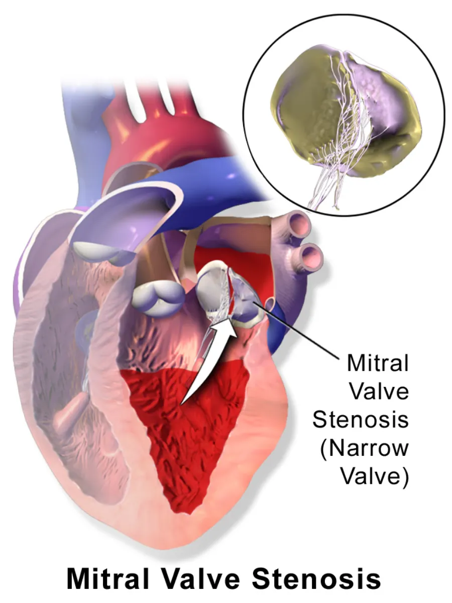
What is mitral valve stenosis?
It most often results from rheumatic heart disease. The left atrium enlarges, predisposing to atrial fibrillation and clot formation.
Mitral Valve Stenosis illustrated
Key points to remember
- Most common cause is rheumatic fever following untreated strep infection.
- Narrowed valve backs blood up into the left atrium and lungs, causing pulmonary congestion.
- Classic findings: dyspnea on exertion, fatigue, orthopnea, and a low-pitched diastolic murmur.
- Left atrial enlargement promotes atrial fibrillation and mural thrombus, raising embolic stroke risk.
- Patients are often anticoagulated when atrial fibrillation is present.
- Treatment may include balloon valvuloplasty or surgical valve repair/replacement.
Common causes
- Rheumatic fever #1 cause
- Narrowed mitral valve
Signs & symptoms
- Dyspnea, orthopnea, fatigue
- Diastolic murmur (rumble)
- Hemoptysis, A-fib
Nursing priorities
- Monitor for signs of pulmonary congestion: dyspnea, crackles, orthopnea, hemoptysis.
- Assess heart rhythm and report atrial fibrillation or irregular pulse.
- Administer anticoagulants, diuretics, and rate-control agents as prescribed and monitor effects.
- Pace activity and provide rest to reduce cardiac workload.
- Educate on infective endocarditis prophylaxis before invasive/dental procedures when indicated.
Labs & values
Patient teaching
- Take anticoagulants exactly as prescribed and attend INR monitoring appointments.
- Report palpitations, increased shortness of breath, or coughing up blood.
- Practice good dental hygiene and inform providers of valve disease before procedures.
- Balance activity with rest to avoid overtaxing the heart.
Red flags: report now
- New irregular pulse or atrial fibrillation with sudden neurologic deficit suggests embolic stroke — emergency.
- Hemoptysis, severe dyspnea, or pink frothy sputum indicates pulmonary edema — report now.
- Acute chest pain with shortness of breath may signal systemic embolism.
References
Clinical references for this topic