Dyspnea, decreased or absent breath sounds, and dullness to percussion over the affected area are characteristic.
Before you read
Test yourself first: 3 questions
Question 1 of 3
During palpation of the posterior chest, the nurse asks the client to say ninety-nine and feels increased vibrations over the right lower lobe. This increased tactile fremitus most likely indicates which condition?
Answer: B. Tactile fremitus increases when lung tissue becomes more dense, such as with consolidation in pneumonia, because solid tissue transmits vibration better than air. Fremitus decreases over a pleural effusion, pneumothorax, or hyperinflated emphysematous lung.
Question 2 of 3
A nurse auscultates a low-pitched, grating sound heard during both inspiration and expiration over the lateral chest that does not change with coughing. The client reports pain with breathing. How should the nurse document this finding?
Answer: C. A pleural friction rub is a low-pitched, grating or rubbing sound produced when inflamed pleural surfaces rub together. It is heard during both inspiration and expiration, does not clear with coughing, and is often associated with pleuritic chest pain.
Question 3 of 3
A nurse observes the fluid in the water-seal chamber of a chest drainage system rising and falling with the client's respirations. How does the nurse interpret this finding?
Answer: A. Tidaling, the rise and fall of fluid in the water-seal chamber with respiration, is an expected normal finding that reflects pressure changes in the pleural space. Absence of tidaling may mean the lung has reexpanded or the tubing is obstructed or kinked.
Keep the streak going with more questions like these, each with a full rationale.
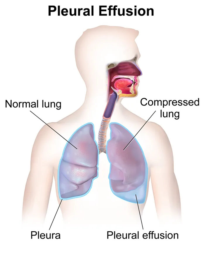
What is pleural effusion?
Transudates result from systemic conditions like heart failure or cirrhosis, while exudates result from inflammation, infection, or malignancy. Large effusions cause dyspnea and require drainage by thoracentesis.
Pleural Effusion illustrated
Key points to remember
- Dyspnea, decreased or absent breath sounds, and dullness to percussion over the affected area are characteristic.
- Decreased chest wall movement and reduced tactile fremitus occur on the affected side.
- Pleuritic chest pain and a dry cough may be present, especially with inflammatory causes.
- A chest x-ray shows blunting of the costophrenic angle and fluid layering.
- Large effusions can cause tracheal shift toward the unaffected side and significant respiratory distress.
- Thoracentesis both relieves symptoms and provides fluid for analysis to determine the cause.
Common causes
- HF, pneumonia, cancer, cirrhosis
Signs & symptoms
- Dyspnea, ↓ breath sounds
- Dull percussion, pleuritic pain
- ↓ chest movement affected side
Nursing priorities
- Position the patient in high Fowler's to ease breathing and assist with thoracentesis.
- Administer oxygen and monitor respiratory status and oxygen saturation.
- Prepare and support the patient for thoracentesis, positioning them sitting up and leaning over a table.
- After thoracentesis, monitor for pneumothorax, assess breath sounds, and observe the puncture site.
- Treat the underlying cause and manage pain to allow effective deep breathing.
Labs & values
Patient teaching
- Report increasing shortness of breath, fever, or chest pain after the procedure.
- Deep breathe and change positions to promote lung expansion.
- Follow up on management of the underlying cause such as heart failure or infection.
Red flags: report now
- Sudden dyspnea, decreased breath sounds, or chest pain after thoracentesis may indicate a pneumothorax.
- Removing more than about 1 liter at once during thoracentesis risks re-expansion pulmonary edema, monitor closely.
- Worsening respiratory distress or tracheal deviation requires immediate escalation.
References
Clinical references for this topic