Sudden severe dyspnea, hypotension, or loss of consciousness signals a massive PE, call for emergency help.
Before you read
Test yourself first: 5 questions
Question 1 of 5
A client with a massive pulmonary embolism is in obstructive shock. Which assessment finding would the nurse most expect?
Answer: A. A massive pulmonary embolism obstructs right ventricular outflow, producing acute right heart strain. Findings include sudden dyspnea, tachycardia, hypotension, hypoxemia, and jugular venous distention from elevated right-sided pressures. Bilateral crackles with frothy sputum are more typical of left-sided failure or pulmonary edema.
Question 2 of 5
A client is admitted with a suspected pulmonary embolism. Which sign or symptom does the nurse most expect to find on assessment?
Answer: B. Sudden onset of pleuritic chest pain with dyspnea and tachypnea is the classic presentation of a pulmonary embolism, often with tachycardia and anxiety. Bradycardia, a productive green cough, and bilateral lower-extremity edema are not characteristic early findings of an acute PE.
Question 3 of 5
A postoperative client suddenly develops dyspnea, chest pain, and tachycardia. The nurse's priority concern is:
Answer: A. Sudden dyspnea, pleuritic chest pain, tachycardia, and hypoxia after surgery/immobility suggest pulmonary embolism — apply oxygen, raise the head of bed, and notify the provider/rapid response.
Question 4 of 5
A client with a DVT is started on anticoagulation. Which nursing action is appropriate?
Answer: B. Do NOT massage the affected leg (embolism risk). Maintain anticoagulation, monitor for bleeding and signs of pulmonary embolism (sudden dyspnea, chest pain), and elevate the leg per orders.
Question 5 of 5
A nurse is reviewing congenital heart defects. Which defect is classified as an acyanotic defect with increased pulmonary blood flow?
Answer: A. A ventricular septal defect (VSD) causes left-to-right shunting, increasing pulmonary blood flow without cyanosis. Tetralogy of Fallot, transposition of the great arteries, and tricuspid atresia are cyanotic defects.
Keep the streak going with more questions like these, each with a full rationale.
What is pulmonary embolism?
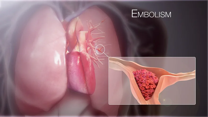
It is a life-threatening emergency because it impairs gas exchange and can cause sudden cardiovascular collapse. Risk factors follow Virchow's triad: venous stasis, hypercoagulability, and endothelial injury (immobility, surgery, pregnancy, oral contraceptives).
Pulmonary Embolism illustrated
Key points to remember
- Sudden onset of dyspnea and sharp pleuritic chest pain are the most common presenting signs.
- Tachypnea, tachycardia, anxiety, and a sense of impending doom are frequent.
- Hypoxemia, cough, and sometimes hemoptysis occur as lung tissue is deprived of perfusion.
- A large saddle embolus can cause hypotension, jugular vein distention, and obstructive shock.
- ABGs often show respiratory alkalosis with hypoxemia and a low PaCO2 from hyperventilation.
- Spiral CT pulmonary angiography is the gold-standard diagnostic test; a positive D-dimer is sensitive but not specific.
Signs & symptoms
- Sudden SOB, sharp chest pain
- Tachycardia, tachypnea, ↓ O₂
- Anxiety, hemoptysis, cough
Nursing priorities
- Apply high-flow oxygen and place the patient in high Fowler's position immediately.
- Notify the rapid response or provider and prepare for anticoagulation.
- Administer anticoagulants (heparin) as ordered, and thrombolytics for massive PE with hemodynamic instability.
- Establish IV access and continuously monitor cardiac rhythm, oxygenation, and vital signs.
- Keep the patient calm and on bed rest to reduce oxygen demand and clot dislodgement.
Labs & values
Patient teaching
- Prevent clots by ambulating early after surgery, doing leg exercises, and staying hydrated.
- Avoid prolonged immobility, take stretch breaks on long trips, and use prescribed compression devices.
- While on anticoagulants, report any unusual bleeding or bruising and attend lab monitoring.
Red flags: report now
- Sudden severe dyspnea, hypotension, or loss of consciousness signals a massive PE, call for emergency help.
- Falling blood pressure with rising heart rate indicates obstructive shock and impending arrest.
- Signs of bleeding during anticoagulant or thrombolytic therapy require immediate evaluation.
- Never massage the legs of a patient with suspected DVT, which can dislodge the clot.
References
Clinical references for this topic