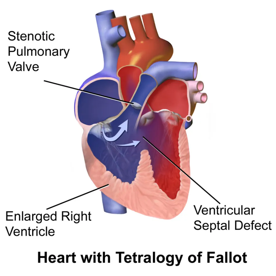
Cyanosis appears because unoxygenated blood is shunted right-to-left across the VSD into systemic circulation.
Before you read
Test yourself first: 3 questions
Question 1 of 3
A nurse caring for an infant with tetralogy of Fallot observes the infant become acutely cyanotic and irritable during feeding (a hypercyanotic, or tet, spell). Which is the priority nursing action?
Answer: A. During a hypercyanotic spell, placing the infant in the knee-chest position increases systemic vascular resistance and decreases the right-to-left shunt, improving pulmonary blood flow. Supplemental oxygen and calming the infant follow. Lying flat or stimulating the infant would worsen the spell.
Question 2 of 3
A nurse is assessing infants for congenital heart defects. Which defect is classified as a cyanotic heart defect?
Answer: B. Tetralogy of Fallot is a cyanotic defect because deoxygenated blood is shunted right to left into the systemic circulation. Atrial septal defect, ventricular septal defect, and patent ductus arteriosus are acyanotic left-to-right shunts.
Question 3 of 3
An infant with tetralogy of Fallot suddenly becomes acutely cyanotic, irritable, and dyspneic during feeding (a hypercyanotic or tet spell). What is the nurse's priority action?
Answer: B. Placing the infant in a knee-chest position during a hypercyanotic spell increases systemic vascular resistance and venous return, improving pulmonary blood flow and oxygenation. Oxygen and calming the infant are also part of management.
Keep the streak going with more questions like these, each with a full rationale.
What is tetralogy of fallot?
The combined defects cause deoxygenated blood to shunt right-to-left and bypass the lungs, producing cyanosis. Remember the four parts with PROVe: Pulmonary stenosis, Right ventricular hypertrophy, Overriding aorta, VSD.
Tetralogy of Fallot illustrated
Key points to remember
- Cyanosis appears because unoxygenated blood is shunted right-to-left across the VSD into systemic circulation.
- Tet spells (hypercyanotic episodes) cause acute deepening cyanosis, irritability, and dyspnea, often during crying, feeding, or stooling.
- Older infants and toddlers instinctively squat or are placed knee-to-chest to relieve a tet spell by increasing systemic vascular resistance.
- A loud systolic murmur is heard at the left sternal border from pulmonary stenosis.
- Chronic hypoxemia leads to clubbing of fingers and toes and polycythemia.
- Chest X-ray classically shows a boot-shaped heart.
Signs & symptoms
- TET spell: sudden cyanosis, crying
- Clubbing, ↓ O2 sat, murmur
Nursing priorities
- During a tet spell, immediately place the infant in a knee-to-chest position and remain calm.
- Administer oxygen and morphine as ordered during hypercyanotic spells to improve flow and reduce agitation.
- Cluster care and feed in small amounts to minimize crying and oxygen demand; allow frequent rest.
- Monitor oxygen saturation, color, respiratory effort, and for signs of dehydration that worsen polycythemia.
- Prepare the family and child for surgical repair and provide pre- and postoperative care.
Labs & values
Patient teaching
- Learn to recognize tet spells and immediately place the child in a knee-to-chest position and call for help.
- Feed slowly in small amounts and allow rest to reduce crying and oxygen demand.
- Keep the child well hydrated to prevent blood from thickening.
- Keep all cardiology follow-up and surgical appointments and report increasing blue color or fainting.
Red flags: report now
- A tet spell with rapidly deepening cyanosis, limpness, or loss of consciousness is an emergency.
- Worsening cyanosis, syncope, or seizures during a spell require immediate intervention.
- Polycythemia increases stroke risk; report signs of dehydration or neurologic changes promptly.
- Never leave a cyanotic infant in distress; position knee-to-chest and call for help at once.
References
Clinical references for this topic