The affected limb is typically swollen, warm, red, and tender, often calf or thigh.
Before you read
Test yourself first: 3 questions
Question 1 of 3
A nurse notes an area of intact skin over the heel that is a persistent, nonblanchable deep red, maroon, or purple discoloration. The client reports the area is painful and boggy. How does the nurse classify this finding?
Answer: B. A deep tissue pressure injury presents as intact or nonintact skin with a localized area of persistent nonblanchable deep red, maroon, or purple discoloration, or a blood-filled blister, caused by damage to underlying soft tissue. The skin may feel boggy or firm and painful. It differs from Stage 1, which is nonblanchable erythema (red, not purple) without deep-tissue damage.
Question 2 of 3
A nurse identifies a localized area of intact skin that is persistent, nonblanchable, deep red, maroon, or purple, with no other tissue loss. How should this be classified?
Answer: A. A deep tissue pressure injury presents as persistent nonblanchable deep red, maroon, or purple discoloration of intact or blistered skin caused by damage to underlying soft tissue. It can rapidly evolve to reveal a full-thickness wound even with treatment.
Question 3 of 3
A nurse is caring for a client 30 minutes after a vaginal birth. The fundus is firm and midline, but the client reports severe, deep, unrelenting perineal and rectal pain that is not relieved by ice or analgesia, and the perineum appears tense and bluish. What does the nurse suspect?
Answer: A. Severe, unrelenting, deep perineal/rectal pain disproportionate to expected discomfort, with a tense, bulging, discolored (bluish/purple) perineum, is the classic presentation of a perineal or vaginal hematoma from concealed bleeding into the tissues. The firm midline fundus rules out atony. The nurse should notify the provider promptly, as a large hematoma can cause significant blood loss.
Keep the streak going with more questions like these, each with a full rationale.
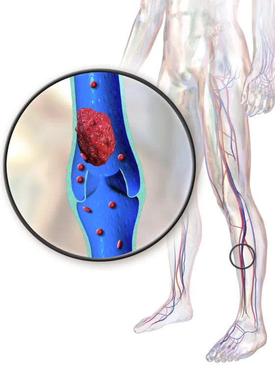
What is deep vein thrombosis?
The most dangerous complication is pulmonary embolism, which occurs if the clot dislodges and travels to the lungs. Risk factors include immobility, surgery, pregnancy, oral contraceptives, and cancer.
Deep Vein Thrombosis illustrated
Key points to remember
- The affected limb is typically swollen, warm, red, and tender, often calf or thigh.
- Unilateral leg swelling with calf pain is a classic finding.
- Virchow's triad of venous stasis, endothelial injury, and hypercoagulability explains clot formation.
- Pulmonary embolism is the major life-threatening complication if the clot embolizes.
- Risk increases with immobility, recent surgery, long travel, pregnancy, estrogen use, and malignancy.
Signs & symptoms
- Unilateral leg swelling, warmth
- Redness, calf pain, tenderness
Nursing priorities
- Maintain bed rest as ordered initially and elevate the affected extremity to reduce swelling.
- Administer anticoagulant therapy such as heparin or low-molecular-weight heparin as prescribed.
- Apply warm compresses if ordered and avoid massaging or rubbing the affected leg.
- Apply sequential compression devices to unaffected limbs and encourage early ambulation for prevention.
- Monitor for signs of pulmonary embolism and for bleeding from anticoagulation.
Labs & values
Patient teaching
- Take anticoagulants exactly as prescribed and watch for and report any signs of bleeding.
- Avoid prolonged sitting or standing, ambulate frequently, and stay hydrated, especially during long travel.
- Do not cross the legs or wear constrictive clothing, and report new leg swelling, sudden chest pain, or shortness of breath.
Red flags: report now
- Sudden dyspnea, sharp pleuritic chest pain, tachycardia, or hypoxia signals pulmonary embolism and is a medical emergency.
- Never massage or rub the affected limb because it can dislodge the clot.
- Signs of anticoagulation bleeding such as hematuria, black stools, or unusual bruising must be reported immediately.
References
Clinical references for this topic